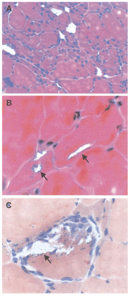
FIG. 6.
Muscle biopsy analysis. Figure 5A is an H&E stain which illustrates the myopathic features with variation in fiber size and grouped regions of muscle fiber atrophy. Figure 5B is also an H&E stain which shows blue-rimmed vacuoles in small and large muscle fibers. Figure 5C is stained with Congo red and illustrates blue rimmed vacuoles with punctate, blue staining debris.

