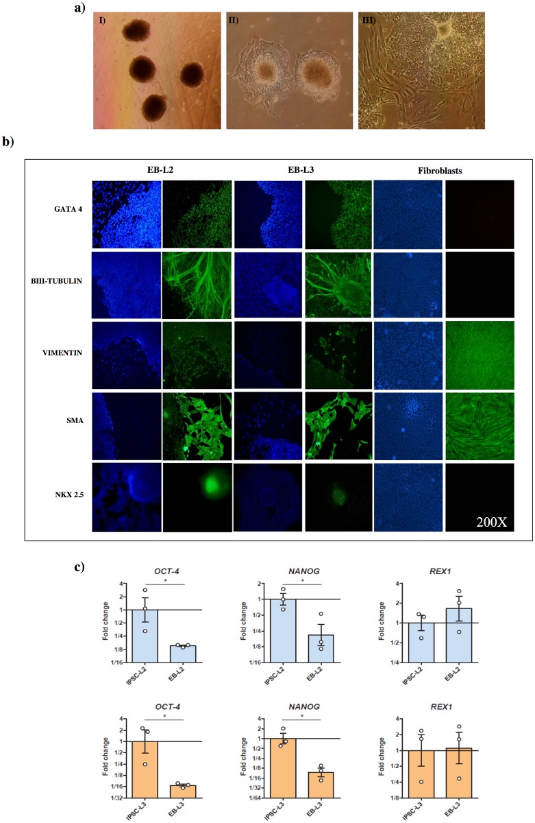
Fig 3. In vitro differentiation of L2 and L3 iPSCs to embryo bodies (EB).
a) Generation of EBs, I) EBs in suspension for 1 week in DMEM medium, II) EBs after 3 days in adherent dishes, III) EBs after 2 weeks in adherent dishes; b) Inmmunofluorescence of endodermal, ectodermal and mesodermal markers in EBs derived from L2 (EB-L2), L3 (EB-L3) iPSCs lines and fibroblasts as controls. In blue nucleous are stained with DAPI c) RT-qPCR analysis of pluripotent markers in L2 vs. EB-L2 and L3 vs. EB-L3. Results are presented as means ± SEM (n = 3). Data were relativized to L2 or L3 for EB-L2 and EB-L3, respectively. *Statistically different (p<0.05).

