Fig. 4.

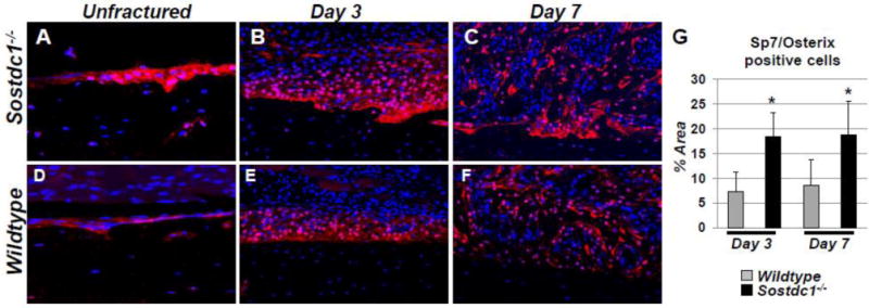
SP7/Osterix (Osx) osteoblast precursor levels are dramatically increased in Sostdc1−/− mice during fracture repair. Immunostains for Osx in Sostdc1−/− mice show an increased number of positive cells at the resting periosteal bone surface of unbroken bones compared to WT controls (A, D). The layer of cells that participate in the periosteal reaction at D3 post fracture is thicker and has more Osx-positive cells, especially at the periosteal surface, compared to controls (B, E). At D7 post-fracture, Osx levels are activated in developing intramembranous callus, which is larger and contains more positive cells in Sostdc1−/− mice, especially at the periosteal surface (C, F). Quantitation of signal area at D3 and D7 revealed an increased area of Osx-positive cells in Sostdc1−/− mice at both time points examined (G).
