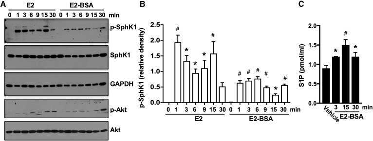
Fig. 3.
Membrane-impermeable E2-BSA activates SphK1 and enhances secretion of S1P. A, B: MDA-MB-231 cells treated with E2 (100 nM) or with E2-BSA (100 nM) for the indicated times. A: Proteins in cell lysates were separated by SDS-PAGE and immunoblotted with the indicated Abs. B: Phospho-SphK1 was quantified by densitometry, and data are expressed as relative density of p-SphK1 normalized to SphK1. C: Cells from duplicate MDA-MB-231 cultures were treated without or with E2-BSA (100 nM) for the indicated times. S1P released into the medium during 30 min secretion assays was measured by LC/ESI/MS/MS. Data are mean ± SD. * P ≤ 0.05; # P < 0.001 (compared with control).

