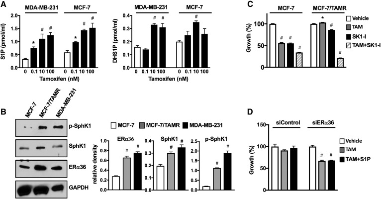
Fig. 7.
Tamoxifen stimulates S1P production, and tamoxifen resistance is associated with increased ERα36 and SphK1 activation. A: MDA-MB-231 and MCF-7 cells were treated without or with the indicated concentrations of tamoxifen for 30 min. S1P and DHS1P released into the medium were measured by LC/ESI/MS/MS. Data are mean ± SD. * P ≤ 0.05. B: Expression levels of SphK1, p-SphK1, and ERα36 were determined by immunoblot analysis in MDA-MB-231, MCF-7/TAMR-1, and parental MCF-7/S0.5 cells. Blots were stripped and reprobed with anti-GAPDH Ab to show equal loading and transfer. Indicated proteins were quantified by densitometry, and data are expressed as relative densities normalized to GAPDH. A, B: * P ≤ 0.05, #P < 0.001 (compared to vehicle). C: MCF-7/TAMR-1 and MCF-7/S0.5 cells were treated with vehicle, tamoxifen (10 µM), SK1-I (15 µM), or both for 2 days, and cell growth was determined. D: MCF-7/TAMR-1 cells transfected with control siRNA or with siRNA targeted to ERα36 (#3) were treated with vehicle or tamoxifen (1 µM) in the absence or presence of S1P (100 nM) for 2 days, and cell growth was determined. C, D: Data are expressed as percent of vehicle-treated control and are mean ± SEM. * P ≤ 0.05; # P < 0.001 (compared with vehicle).

