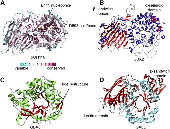
Fig. 3.
GH1, GH59, and GH116 general fold topology. A: G116 TxGH116 structure (PDB ID: 5bvu) (57) with the position of the conserved amino residues of GH116 members depicted on the TxGH116 structure based on sequence alignment using the ConSurf Server. B: GBA2 model based on the TxGH116 structure. C: GH1 glycosylceramidase GBA3 structure (PDB ID: 2e9m) (66). D: GH59 GALC (PDB ID: 4ccc) (73).

