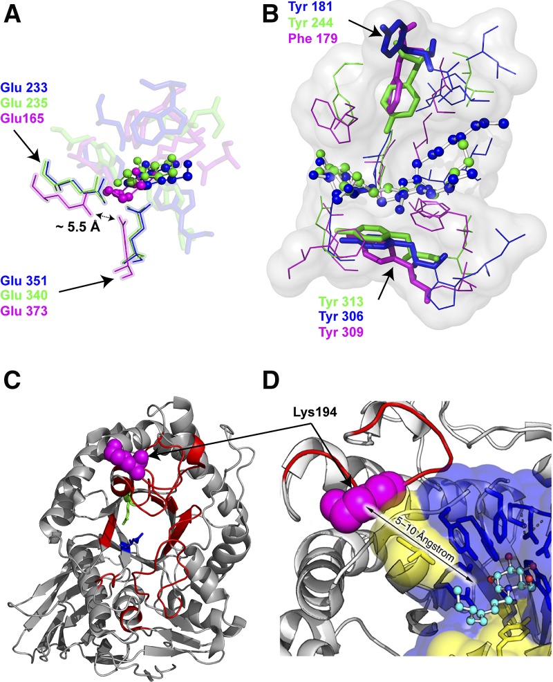
Fig. 4.
Glycosylceramidases: binding site architecture and stabilization. A: Superposition of the glycon binding sites of EGC II [PDB ID: 2osx (37); blue], GBA [PDB ID: 2v3e (83); green], and GBA3 [PDB ID: 2e9m (66); magenta] in complex with d-glucose (ball-and-stick, blue), DNJ (ball-and-stick, green) and galactose (ball-and-stick, magenta), respectively. B: Superposition of the aglycon sites of EGC II in complex with glucosylceramide (ball-and-stick, blue), GBA1 in complex with NN-DNJ (ball-and-stick, green), and GBA3 (ball-and-stick, magenta). The conserved aromatic residues are shown in sticks. C: GBA1-rigidified regions upon IFG bonding depicted in red on the GBA1 crystal structure (PDB ID: 2v3e) as determined by HDX mass spectrometry (87). D: Tryptic cleavage site (Lys 194) highlighted in magenta spheres on the GBA1 structure (PDB ID: 1ogs) (33).

