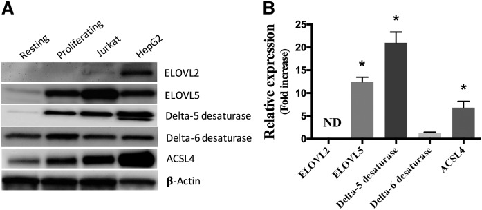
Fig. 3.
Protein expression of indicated enzymes. Proteins (10 μg) from primary resting and proliferating T-cells, Jurkat cells, and HepG2 cells were separated on 10% SDS-PAGE gels and transferred onto a PVDF membrane. Western blotting was performed using primary and secondary antibodies as listed in the Material and Methods section. A: Western blots representative of three independent experiments. B: The relative expression of proteins in proliferating T-cells compared with resting T-cells was determined by densitometry using β-actin for normalization. The values are means ± SEMs of three independent experiments. For T-cells, each independent experiment was conducted with cells obtained from a different subject. *Different from resting cells (P < 0.05) as determined by paired two-sided Student’s t-test. ND, not determined.

