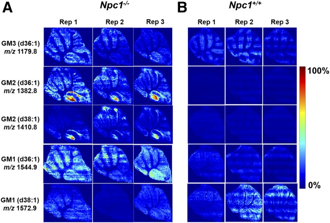
Fig. 2.
MALDI-MS images of major gangliosides found to be increased in 7-week-old Npc1−/− (A) and Npc1+/+ (B) cerebellum with serial sections for each mouse at a pitch of 50 μm. Substantial accumulation is detected for GM3 (d36:1) (m/z 1,179.8) in the Purkinje layer, specifically in lobule X, whereas the accumulation of GM2 (d36:1) (m/z 1,382.8) and GM2 (d38:1) (m/z 1,410.8) is determined to be localized in lobule X. GM1 (m/z 1,544.9) does not reveal localization. On the other hand, GM1 (m/z 1,572.9) does not appear to be accumulating or localizing in the KO mouse compared with the WT. Triplicate analysis shows reproducible ion intensities and lipid distributions, and images were normalized by the TIC of each section using MSiReader.

