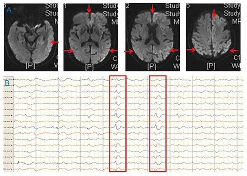
Figure 1.

MRI (A) findings of bilateral cortical diffusion restriction presenting with hyperintensive signal in the cortex on diffusion weighted imaging. EEG (B) showed slowing and periodic discharges over bilateral hemisphere with triphasic morphology.
