. 2018 Dec 3;7:e27706. doi: 10.7554/eLife.27706

Figure 1. SOX4 is a transcriptional activator, preferentially binding to open/active chromatin.

(A) Venn-diagram showing the overlap of SOX4 binding sites identified by ChIP-seq in WT-HMLE and DOX-induced HMLE-S4 cells. (B). Genomic distribution of SOX4 binding sites in WT-HMLE cells with respect to annotated genes, represented as distance from the nearest TSS. Random genomic regions were used as background. (C). De novo motif analysis of SOX4-bound sites in DOX-treated HMLE-S4 cells. Selected significantly enriched motifs are represented. SOX4 was identified as the most highly enriched motif. (D) Density of the consensus SOX4 motif in SOX4 bound sites in HMLE-S4 cells 500 bp up- and downstream of the peak center. (E) Occupancy plots of SOX4, H3K27ac, H3K27me3, POL2 and H3K4me3 in the 10kb-genomic region surrounding the SOX4 peak center. (F) Changes in SOX4, H3K27ac, H3K4me3, POL2 and H3K27me3 are shown for SOX4-bound and SOX4-unbound sites (5%–95% whiskers, two-tailed Mann-Whitney U test, **** indicate p<0.0001). (G) Changes in SOX4, H3K27ac, and POL2 in HMLE-S4 for top 1000 DOX-induced sites ranked by SOX4-signal (****p<0.0001, Wilcoxon-signed-rank test) (H) Genomic tracks representing the occupancy of SOX4, H3K27me3, H3K4me3, H3K27ac and POL2 in untreated and DOX-treated HMLE-S4 cells surrounding the genomic locus of the canonical SOX4 target gene TEAD2.

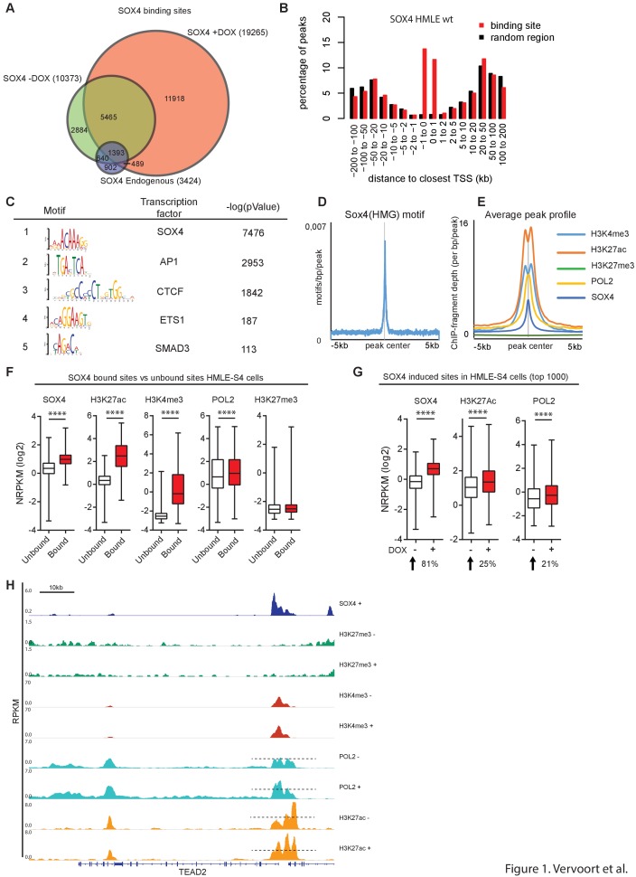
Figure 1—source data 1. Analyses of Rapid Immunoprecipitation for Mass spectrometry on Endogenous proteins (RIME) to identify an optimaal anti-SOX4 antibody for ChIP described in Figure 1A.
DOI: 10.7554/eLife.27706.004
Figure 1—source data 2. Overview of ChIPseq and RNAseq experiments described in Figures 1 and 2.
DOI: 10.7554/eLife.27706.005

Figure 1.

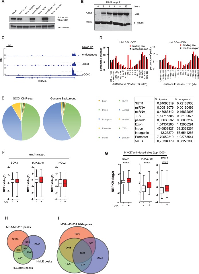
Figure 1—figure supplement 1. Antibody validation and ChIP-seq analysis.

Figure 1—figure supplement 1.

(A) Commercially available and a custom-made SOX4 antibodies were tested for their ability to immunoprecipitate (IP) HA-tagged SOX4. HEK293T cells were transiently transfected with HA-tagged SOX4. Two days post transfection, cells were lysed in radio-immunoprecipitation buffer (RIPA) and IPs were formed using the indicated antibodies including an antibody against the HA-epitope. This demonstrated that the SOX4 diagenode antibody was most efficient at immunoprecipitating SOX4. (B) Validation of the successful lentiviral transduction and inducibility of the DOX-inducible pINDUCER-Sox4 construct in HMLE-cells. Transduced cells were treated with DOX (1 µg/ml) for the indicated time points after which induction of SOX4 expression was analyzed using an anti-HA antibody. (C) A representative example of SOX4 occupancy profiles derived by ChIP-seq normalized for sequencing depth (reads per kilobase per million reads sequenced; RPKM) in wt-HMLE and non-induced and induced HMLE-S4 cells. The genomic regions surrounding the HDAC2 locus is shown. (D) Histogram representing the distance of SOX4 peaks relative to the TSSs of annotated genes. (E) Genomic distribution of SOX4 peaks in DOX-treated HMLE-S4 cells over annotated regions compared to the genomic background. (F–G) Quantitative analysis of enhancer regions as defined by H3K27ac peaks. Normalized read density used to determine the sites without or with DOX-induced changes in SOX4, H3K27ac, H3K4me3, POL2 and H3K27me3 occupancy. Data are shown as boxplots with 5–95% whiskers. Statistical comparison was performed with a two-tailed Wilcoxon-signed-rank test (****p<0.0001) (H) Venn-diagram representing the overlap of SOX4 binding sites in MDA-MB-231, HCC1954 and DOX-treated HMLE-S4 cells. (I) Venn-diagram showing the overlap between genes with a SOX4 peak within 20 kb of their TSS in MDA-MB-231, HCC1954 and DOX-treated HMLE-S4 cells.