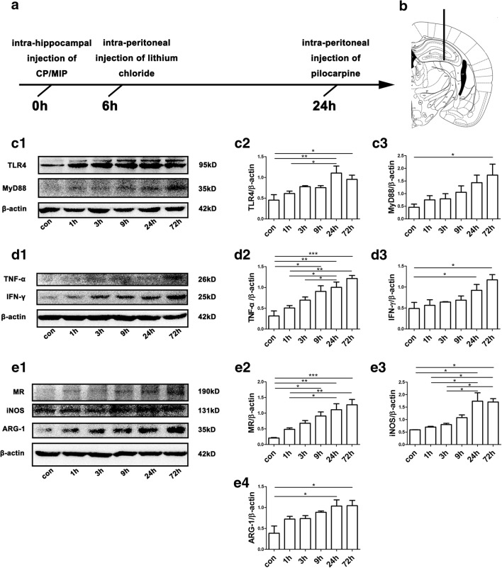
Fig. 1.
Experimental design and protein levels of markers for M1/M2 polarization and TLR4–MyD88 signaling in mouse hippocampi across time points in the acute phase after SE. (A) Schematic illustration of the experimental procedure. CP or MIP was injected intrahippocampally 6 h prior to an intraperitoneal injection of lithium chloride. Twenty-four hours after the intrahippocampal injection, pilocarpine was administered intraperitoneally. SE was halted by diazepam administration. The mice were sacrificed at different time points after SE, and their brains were collected for analysis. (B) Site of stereotactic unilateral hippocampal injection. (C1) Representative immunoblots illustrate that the abundance of TLR4 and MyD88 in mouse hippocampi increased over time in the acute phase after SE. (C2–C3) Group comparison of TLR4 and MyD88 immunoblots (calibrated relative to β-actin). (D1) Representative immunoblots showing increases in TNF-α and IFN-γ in mouse hippocampi over time up to 72 h after SE. (D2–D3) Group comparison of TNF-α and IFN-γ levels. (E1) Representative immunoblots of MR, iNOS, and ARG-1 for each group; note the relatively rapid increase in iNOS levels over time. (E2–E4) Comparison of MR, iNOS, and ARG-1 levels among the above groups. N = 6 per group in the Western blots; *p < 0.05, **p < 0.01, ***p < 0.001 between groups; ANOVA followed by Tukey’s test

