Fig. 6.
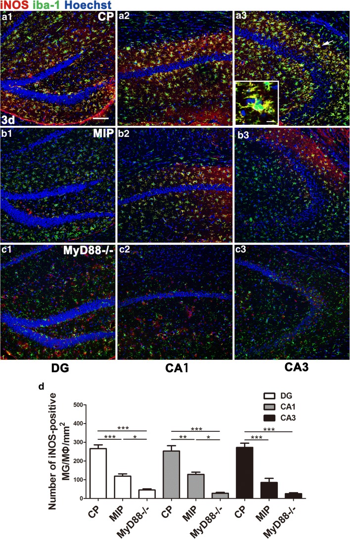
Effect of MyD88 inhibition and MyD88 deficiency on the numbers and distribution of M1 MG/MΦ in the hippocampus 3 days after SE. Immunofluorescence microscopy showed iNOS-positive MG/MΦ in the DG, CA1, and CA3 of mice in the CP group (A1–A3), MIP group (B1–B3), and MyD88−/− group (C1–C3) at 3 days. The inset of (A3) shows a high-magnification view of iNOS and iba-1 double-labeled cells (indicated by arrows) in the CA3. The colocalization of iNOS and iba-1 indicated in yellow shows that iNOS labeling appears in nearly the entire MG/MΦ, including the processes and somata. (D) Group comparison of the numbers of iNOS/iba-1 double-positive cells in the DG, CA1, and CA3 (means ± SEM, n = 3). *p < 0.05, **p < 0.01, ***p < 0.001 between groups; 1-way ANOVA followed by Tukey’s test. Scale bars: (A1–C3) 100 μm; (A3) (inset) 12.5 μm

