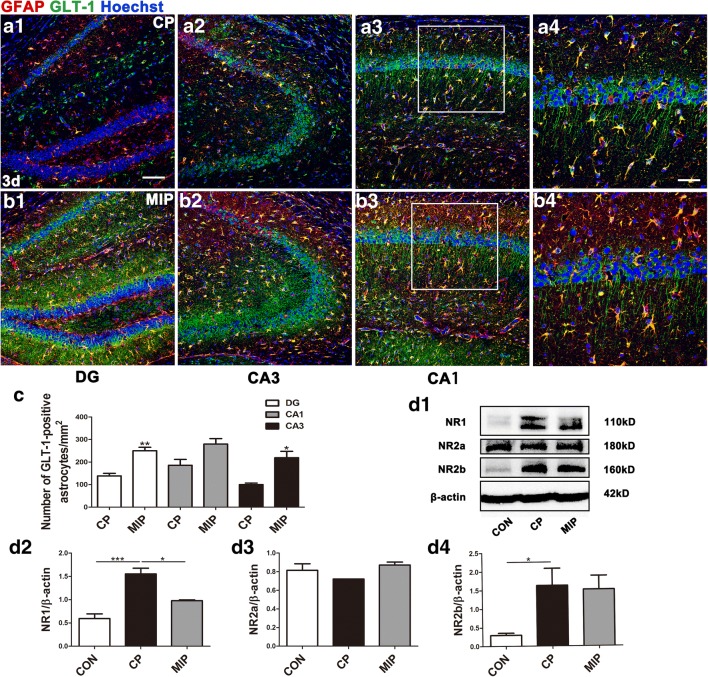
Fig. 9.
Hippocampal GLT-1 and NR1 expression 3 days after SE with MyD88 inhibition. Sections from the hippocampi of mice in the CP group (A1–A3) and MIP group (B1–B3) 3 days after SE with GLT-1 immunoreactivity in astrocytes and neuronal processes. (A4, B4) Higher magnification of the boxes in (A3) and (B3). (C) Comparison of the numbers of GFAP/GLT-1 double-labeled cells in the DG, CA1, and CA3 between the CP and MIP groups (means ± SEM, n = 3). *p < 0.05 versus the CP group; **p < 0.01 versus the CP group. Independent samples t tests were performed. (D1) Immunoblots of NR1, NR2a, and NR2b for the control, CP, and MIP groups. (D2–D4) Comparison of NR1, NR2a, and NR2b levels among the above groups (calibrated to β-actin). *p < 0.05; ***p < 0.001 between groups. One-way ANOVA followed by Tukey’s test. Scale bars: (A1–A3, B1–B3) 100 μm; (A4, B4) 50 μm

