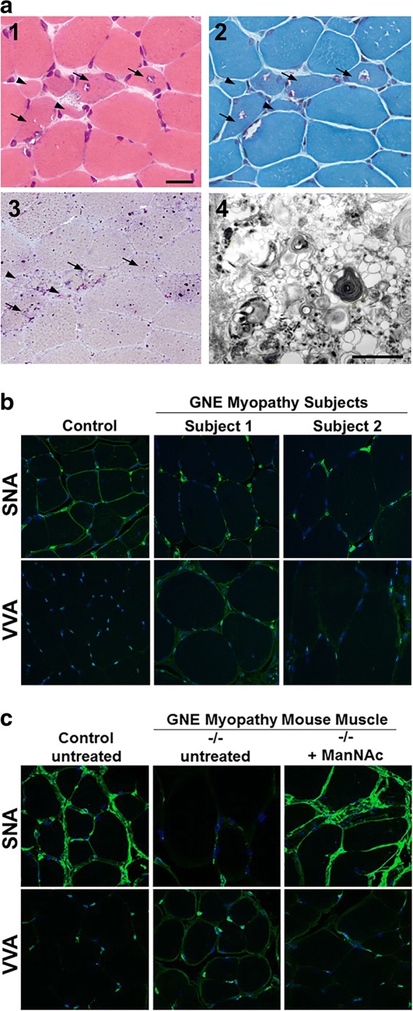
Fig. 4.

GNE Myopathy muscle histology and lectin histochemistry. (a) Stained cryosections of a muscle biopsy (biceps brachii) from a GNE myopathy patient. (1) H&E staining shows variation in muscle fiber size, atrophic fibers (arrowheads), and characteristic fibers with rimmed vacuoles (arrows). (2) Modified Gomori trichrome staining in which rimmed vacuoles appear as vacuoles rimmed by red granules. (3) Acid phosphatase staining showing intense lysosomal staining in regions with rimmed vacuoles, implying that these rimmed vacuolar areas maybe clusters of autophagic vacuoles. (4) Electron microscopy of a rimmed vacuole containing cytoplasmic region, confirming that structures are indeed composed of autophagic vacuoles and myeloid bodies, in addition to excessive cellular debris. Scale bar in (1) represents 50 μm in section images shown in (1, 2, 3). Scale bar in (4) denotes 1 nm. (b, c) Paraffin embedded skeletal muscle sections of control and GNE myopathy subjects (b) and mice (c) stained with the lectins (green) SNA (predominantly binding terminal α(2,6)-linked sialic acid on all glycans) or VVA (predominantly binding terminal GalNAc, without sialic acid attached, O-linked to serine or threonine residues of glycoproteins), costained with the nuclear dye DAPI (blue). GNE myopathy sections show hyposialylation, indicated by decreased staining of SNA and increased staining of VVA, compared to control sections. Muscle sections of GNE myopathy mutant mice (−/−) that received oral ManNAc therapy exhibited a sialylation status restored to the normal range. All confocal imaging was performed at the same microscope intensity settings per tissue and per lectin (with a 63× objective). All images are 1D projections of confocal Z-stacks. Images in (a): adapted with permission from Huizing et al., OMMBID [11]. Images in (b): adapted with permission from: Leoyklang et al. [65]. Images in (c): adapted with permission from Niethamer et al. [68]
