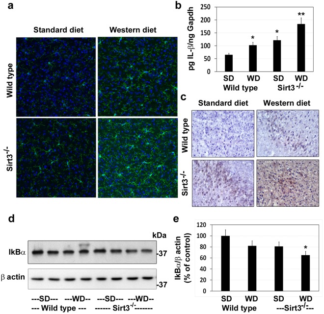
Figure 8.
Microgliosis and markers of neuroinflammation in the brain of western diet-fed Sirt3−/− mice. Wild type and Sirt3−/− mice were fed on standard (SD) and western diet (WD) for four months. (a) Brain samples were subjected to immunofluorescent staining of Iba1 with FITC (green) for microglia. The nuclei were stained with DAPI (blue). Representative images, captured at 400X magnification in a Leica confocal microscope, are presented. (b) The mRNA of IL-1β was determined by real-time PCR in an ABI Prism 7700 sequence detector (Applied Biosystems, Foster City, CA). (c) Immunohistochemical analysis of IL-1β was performed with the mouse brain samples. Representative images are presented. (d) Western blot analysis of IkBα was performed with the mouse brain samples. The blots were reprobed with β actin antibody. (e) The band intensity of IkBα was quantitated by densitometric scanning and corrected for the levels of β actin. The values are mean ± SE of six observations. *P < 0.01; **P < 0.001 vs wild type mice on standard diet.

