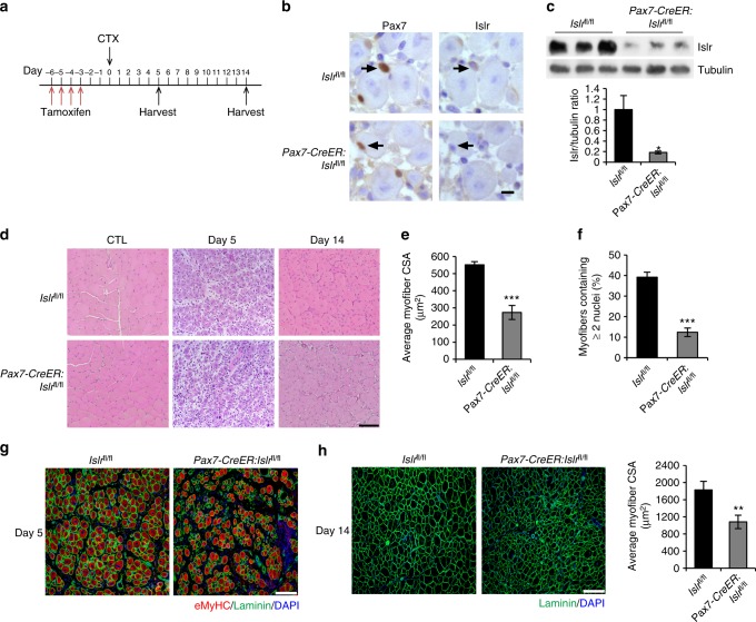
Fig. 5.
Inducible deletion of Islr in satellite cells impairs skeletal muscle regeneration. a Schematic outline of CTX injection in tamoxifen-treated Islrfl/fl and Pax7-creER:Islrfl/fl mice at the age of 10 weeks. b Immunohistochemistry analysis of Islr in Pax7+ cells of Islrfl/fl and Pax7-creER:Islrfl/fl mice at 5 d postinjury using serial sections. Scale bar = 10 μm. c Western blot analysis of Islr protein levels in injured TA muscles of Islrfl/fl and Pax7-CreER:Islrfl/fl mice at 5 d post injury. d H&E staining of injured and CTL in Islrfl/fl and Pax7-CreER:Islrfl/fl mice at 5 and 14 d postinjury. N = 3 in each group. Scale bar = 100 μm. e CSA of regenerating myofibers and f the number of myofibers containing two or more centrally located nuclei per field at 5 d postinjury. N = 3 in each group. g Immunofluorescence analysis of eMyHC+ fibers in TA muscles of Islrfl/fl and Pax7-CreER:Islrfl/fl mice at 5 d postinjury. N = 3 in each group. Scale bar = 100 μm. h Immunofluorescence analysis of the size of myofibers in TA muscles of Islrfl/fl and Pax7-CreER:Islrfl/fl mice at 14 d post injury. N = 3 in each group. Scale bar = 200 μm. The CSAs are shown on the right. Error bars represent the means ± s.d. *P < 0.05, **P < 0.01, ***P < 0.001; Student’s t test

