Fig. 1.
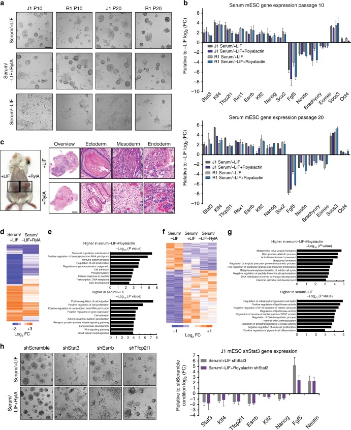
Royalactin maintains stemness in murine embryonic stem cells. a Representative images of J1 and R1 mESCs cultured in serum/+LIF, serum/−LIF, or serum/−LIF + Royalactin for 10 and 20 passages. After LIF withdrawal, mESCs rapidly differentiated, whereas cells cultured with Royalactin supported self-renewal with negligible differentiation. Scale bar, 200 μm. b Quantitative expression of pluripotency and differentiation-associated genes from a. Data are means ± SD (n = 2). c Mice bearing mESC-derived teratomas from J1 mESCs cultured three passages in +LIF and −LIF + Royalactin demonstrated retained pluripotency, and on high magnification (400×) produced differentiated ectodermal, mesodermal, and endodermal tissues. Scale bar, 80 μm. d RNA-seq log2-fold change values in transcript level of all genes in serum/+LIF or serum/–LIF + Royalactin J1 mESCs (passage 10) relative to serum/–LIF. e GO term analysis of differentially expressed genes from d. f ATAC-seq activity in J1 mESCs at passage 10. Each column is a sample, each row is an element. Samples and elements are organized by unsupervised k-means clustering. g GO term analysis of differentially accessible regions from f. h Representative images of Stat3, Esrrb, and Tfcp2l1 knockdown in J1 mESCs with serum/+LIF and serum/−LIF + Royalactin conditions and qPCR analysis of pluripotency and differentiation-associated genes from the same cells. Data are means ± SD (n = 2). Scale bar, 200 μm. RylA Royalactin

