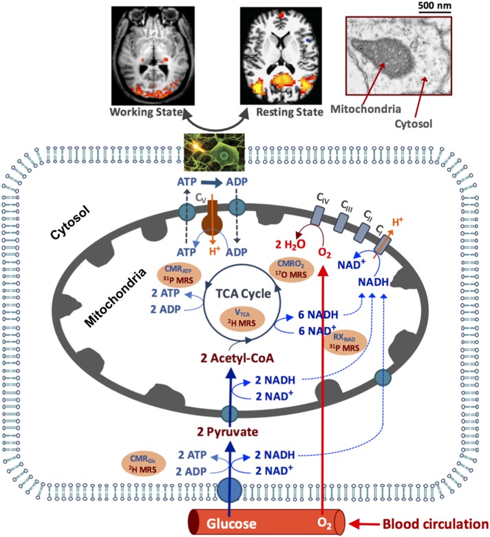
Figure 1.
Schematic illustration of major brain hemodynamic and metabolic processes occur in capillary, cytosol and mitochondrial sub-cellular compartments. The feeding arteries supply oxygen and glucose to the cell; glucose is converted to pyruvate, which enters mitochondrial tricarboxylic acid (TCA) cycle and oxidative metabolism pathways. The oxygen utilization is generally coupled with the adenosine triphosphate (ATP) production via the oxidative phosphorylation of adenosine diphosphate (ADP). The ATP is utilized in cytosol to support electrophysiological activities and brain functions at resting and/or working state. The NAD redox reactions are essential in regulating the ATP energy production. The cerebral metabolic rates of glucose (CMRGlc), oxygen (CMRO2) and ATP production (CMRATP), TCA cycle rate (VTAC) and nicotinamide adenine dinucleotide (NAD) redox ratio (RXNAD) representing the metabolic activities of the brain tissue can be noninvasively measured or imaged using the advanced in vivo X-nuclear 2H, 17O and 31P MRS approaches as depicted in the shadowed texts with orange background. CI-CV represent five enzyme complexes involving in the cellular respiration chain reactions.

