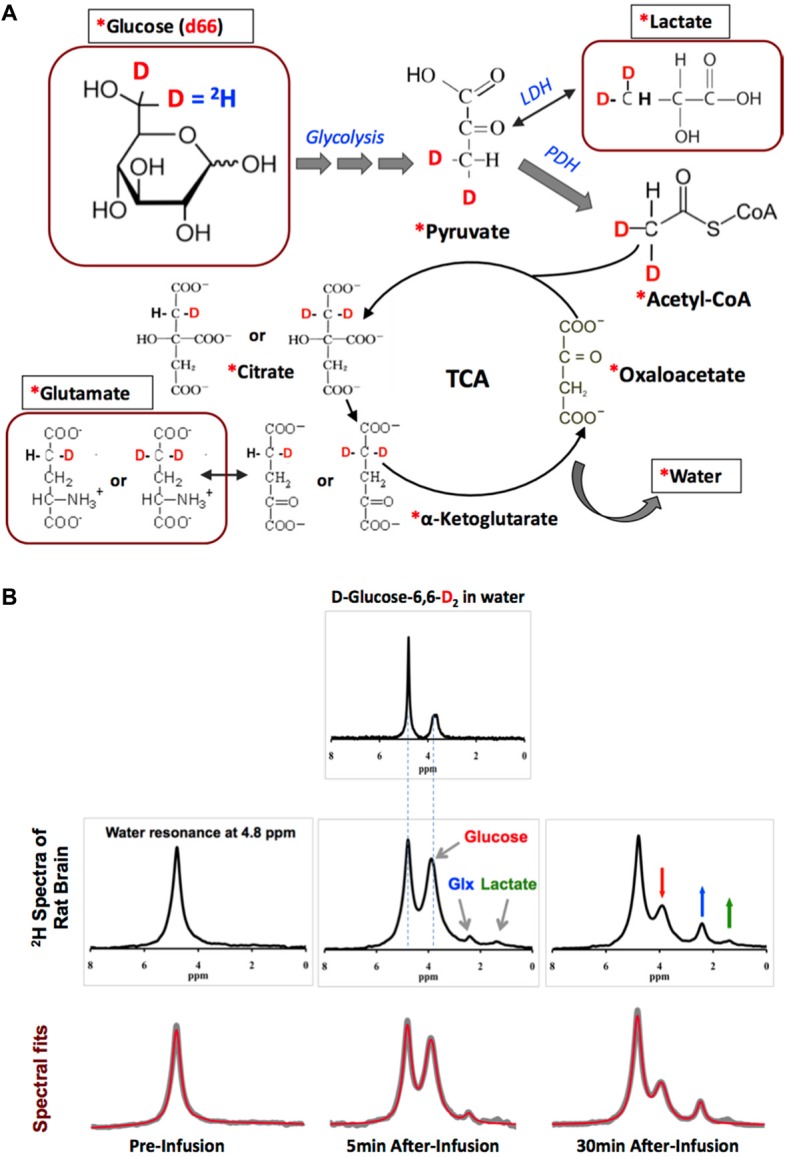
Figure 2.

(A) The 2H-labeling scheme following the metabolic pathways of isotope-labeled glucose D-Glucose-6,6-d2 (d66). The 2H-labeled glucose first incorporates into pyruvate pool through glycolysis to form [3,3-d2] pyruvate, some of which can be converted to [3,3-d2] lactate by lactate dehydrogenase (LDH). [3,3-d2] Pyruvate can also be transported into the mitochondria to form [2,2-d2] Acetyl-CoA catalyzed by pyruvate dehydrogenase (PDH). After entering the TCA cycle, intermediates (4-d) or [4,4-d2] citrate and (4-d) or [4,4-d2] α-ketoglutarate could exchange with glutamate to generate (4-d) or [4,4-d2] glutamate. In this process, the 2H-labels may exchange with the proton(s) in water molecule to form deuterated water and depart from the cycle. “*”: Pools labeled with 2H; square boxes: highlighting the metabolites detectable by in vivo 2H MRS. (B) Representative original (upper rows black traces and bottom row gray traces) and fitted (red traces in bottom row) 2H spectra obtained from deuterated glucose (d66) phantom solution (top panel), and in rat brain pre- (left column) and 5 or 30 min post-deuterated glucose (d66) infusion. 2H resonance assignments: water at 4.8 ppm (use as a chemical shift reference); glucose at 3.8 ppm; mixed glutamate and glutamine (Glx) at 2.4 ppm; and lactate at 1.4 ppm. Figure adapted from Lu et al. (2017).
