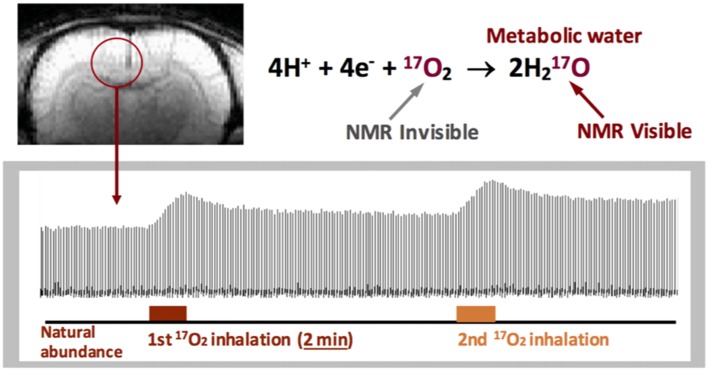
Figure 3.
Stack plots of rat brain tissue H217O spectra obtained from a single voxel selected from the 3D 17O MRSI datasets with two consecutive 17O2 inhalations (~2 min each) for repeated CMRO2 and cerebral blood flow (CBF) imaging measurements. 17O2 in either blood or brain tissue is NMR invisible. Figure adapted from Zhu et al. (2007).

