
Figure 3.
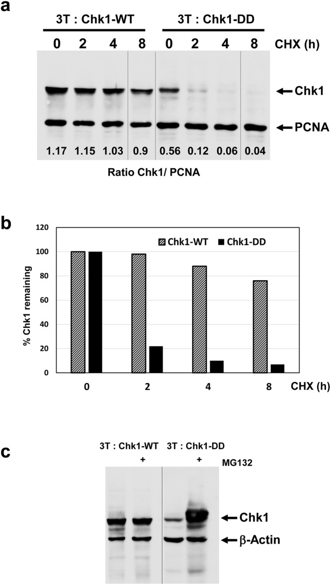
Chk1-DD is subject to rapid proteasomal degradation. (a) 3T: Chk1-WT and 3T: Chk1-DD DT40 cell lines were treated with DOX for 16 hours to induce Chk1 protein expression and then further treated with cycloheximide (CHX) to block protein synthesis for the indicated times. Cell extracts were prepared, 30 μg resolved by SDS-PAGE, and analysed by western blotting using the indicated antibodies. Samples were resolved on the same western blot (see Supplementary Information for original images). (b) Quantification of the data shown in (a), the percentage Chk1 protein remaining at each time point was calculated using PCNA to correct for variations in loading. Control values for t = 0 are converted to 100% to facilitate comparison of relative protein stability. (c) 3T: Chk1-WT and 3T: Chk1-DD cell lines were treated for 16 hours with DOX plus or minus MG132 to inhibit protein degradation via the proteasome. Cell extracts were prepared, 30 μg resolved by SDS-PAGE, and analysed by western blotting using the indicated antibodies. Samples were resolved on two separate western blots (see Supplementary Information for original images).