Figure 6.

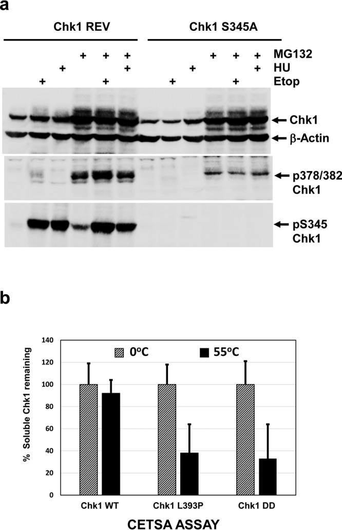
Chk1 T378/382 auto-phosphorylation is not augmented by DNA damage or DNA synthesis inhibition. (a) Chk1 REV DT40 cells stably expressing Chk1-WT, or Chk1-S345A DT40 cells stably expressing a mutant where alanine replaces S345 (see text for additional explanation), were treated for 16 hours with ETOP and HU in the presence or absence of MG132. Cell extracts were prepared, 30 μg resolved by SDS-PAGE, and analysed by western blotting using the indicated antibodies. Samples were resolved on three separate western blots (see Supplementary Information for original images). (b) CETSA analysis of the thermal stability of the Chk1-WT, Chk1 L393P, and Chk1-DD proteins in cell extracts. Protein expression was induced by treatment with DOX for 16 hours and native, soluble whole cell extracts prepared14. Portions of each extract were heated at 55 °C for 5 minutes or retained on ice. Insoluble proteins were then removed by centrifugation and 30 μg resolved by SDS-PAGE in triplicate and analysed by western blotting using antibody recognising total Chk1. Shown is the mean and standard deviation of the percentage of soluble Chk1 remaining after treatment at 55 °C.
