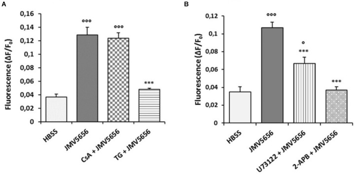
FIGURE 2.
Effects of different inhibitors on JMV5656-mediated intracellular Ca2+ increase in RAW264.7 cells. Cells were loaded with FLUO-4 NW and treated with different inhibitors before the stimulation with 1 μM JMV5656. Graphs show intracellular Ca2+ mobilization (expressed as fluorescence intensity) in RAW264.7 cells stimulated with (A) HBSS (vehicle), JMV5656 alone and JMV5656 in presence of 2 μM CsA (15 min) or 2 μM TG (20 min); and (B) HBSS (vehicle), JMV5656 alone, and JMV5656 in presence of 10 μM U73122 (10 min) or 75 μM 2-APB (15 min). Data are shown as the mean ± SEM of measurements obtained in three independent experiments (n = 18). °p < 0.05, ∘∘∘p < 0.001 vs. HBSS; ∗∗∗p < 0.001 vs. JMV5656 (Tukey’s test).

