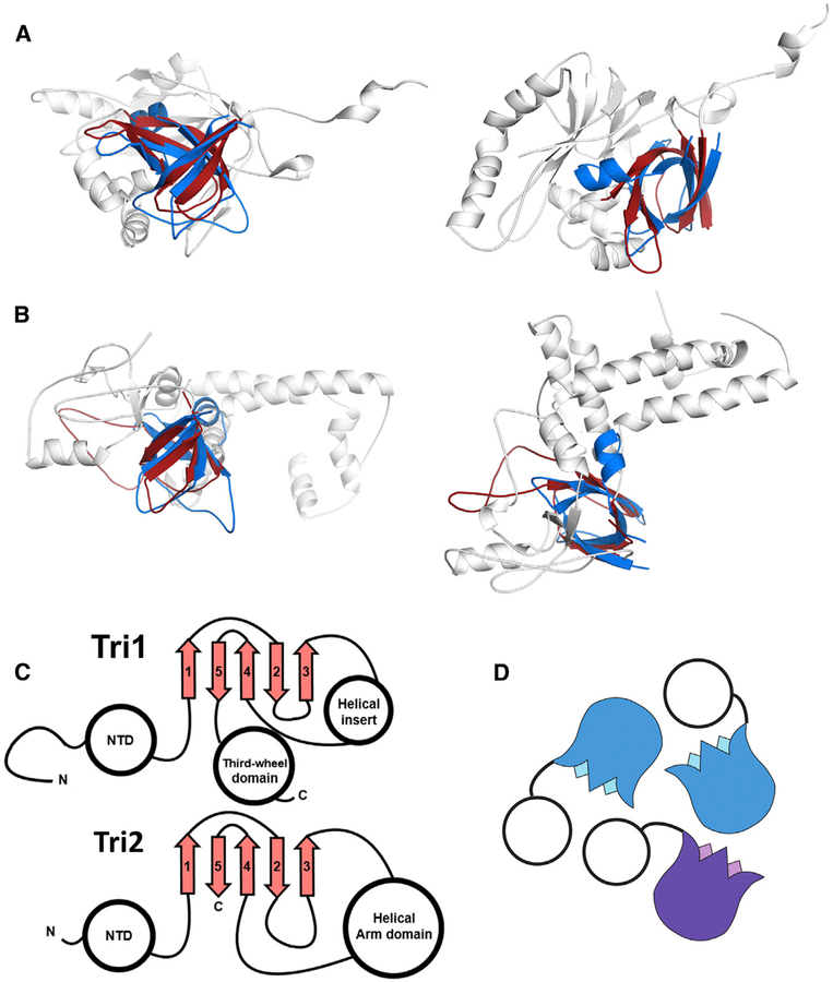
Figure 4. Structural Similarity of Phage Decoration Protein Trimers and the HCMV Triplex.
(A and B) The P74–26 gp87 β tulip domain (blue) is similar to central domain of HCMV Tri1 (A) and Tri2
(B) proteins (gray, β tulip domains in red; PDB: 5VKU).
(C) Central domain of HCMV Tri1 and Tri2 has conserved β tulip topology.
(D) HCMV triplex proteins form an asymmetric trimer consisting of two molecules of Tri2 (blue) and one molecule of Tri1 (purple).

