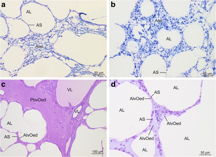
Fig. 3.
Some sections contained areas with microatelectasis or oedema formation. Microatelectasis was seen predominantly in control group (a) or ischaemia group (b). Oedema was found almost only in EVLP groups (c) aEVLP, (d) cEVLP). Light micrographs of toluidine blue stained sections. AL air filled alveolar lumen, AS interalveolar septum, Atel atelectasis, AlvOed intraalveolar oedema, PbvOed peribronchovascular oedema, VL vascular lumen

