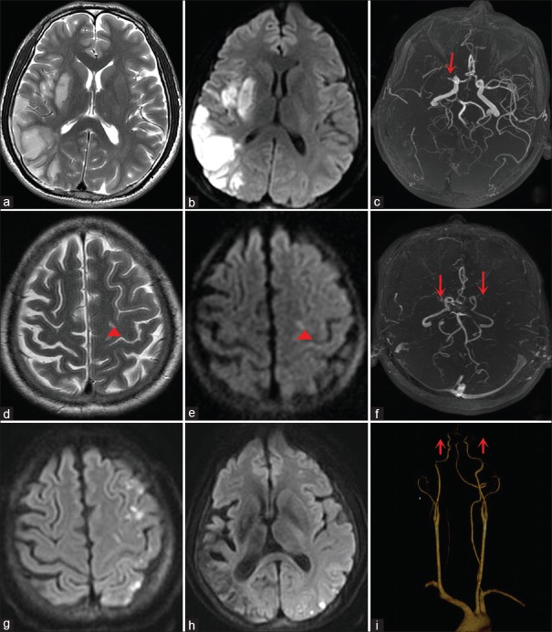
Figure 1.
(a and b) Brain MRI scanned in 2011 showed acute infarction in the right temporal lobe and basal ganglion. (c) MRA showed high-grade stenosis in the right middle cerebral artery (red arrow). (d and e) Brain MRI scanned in 2016 showed punctuate infarction in the left frontal lobe (red short arrowheads). (f) MRA showed severe stenosis in the terminal portion of bilateral internal carotid arteries (red arrows). (g and h) Repeated DWI showed multiple punctate hyperintensity in the left hemisphere. (i) CTA showed severe stenosis in terminal portion of bilateral internal carotid arteries (red arrows). MRI: Magnetic resonance imaging; MRA: Magnetic resonance angiography; DWI: Diffusion-weighted imaging; CTA: Computed tomography angiography.

