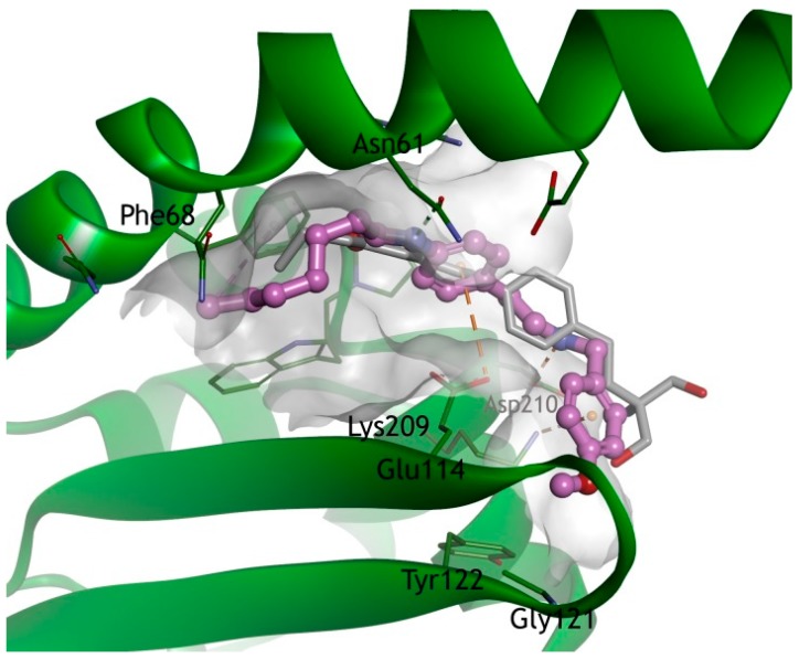
Figure 4.
Docking model of FTY720 (1, gray, ball and stick) and compound 7 (purple, ball and stick) in the I2PP2A/SET. The X-ray structure of IPP2A/SET was obtained from the Protein Data Bank (PDB code 2E50). The I2PP2A/SET is represented by a blue ribbon model. The hydrogen bonds are shown as a green dashed line, and an orange dashed line represents electrostatic interactions. Also, the hydrophobic interactions are shown as a pink dashed line.

