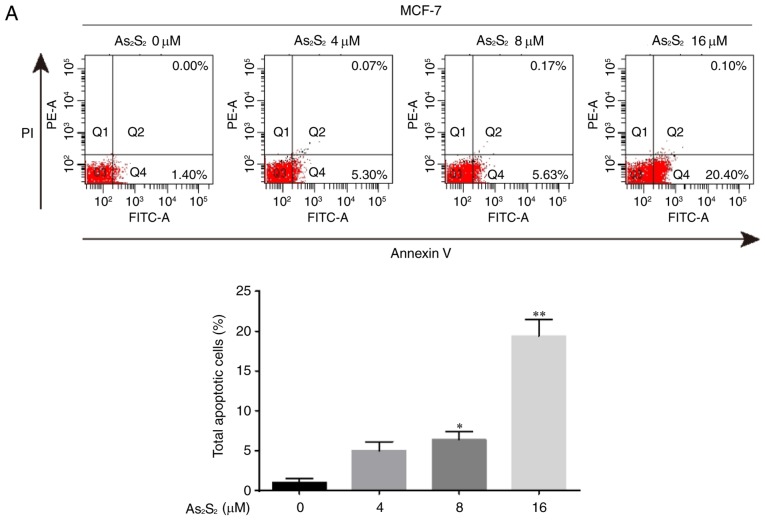
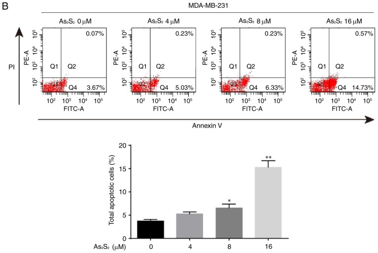
Figure 8.
As2S2 induces apoptosis in breast cancer cells. (A) MCF-7 and (B) MDA-MB-231 cells were treated with different concentrations of As2S2 (0, 4, 8 and 16 µM) for 48 h, followed by staining with Annexin V/PI, and then analyzed by flow cytometry. The cells were assessed for the total number of apoptotic cells, including early-apoptotic (Annexin V+/PI−) and late-apoptotic (Annexin V+/PI+) cells. Results are expressed as the mean ± standard error of the mean (n≥3). *P<0.05, **P<0.01 vs. control (0 µM As2S2). PI, propidium iodide; PE, phycoerythrin.


