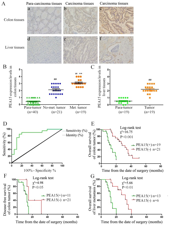
Figure 1.
PEA15 expression in colorectal cancer and metastatic liver cancer tissues and its correlation with clinical prognosis of patients with liver metastasis of colorectal cancer. (A-C) IHC was used to detect the expression of PEA15 in colorectal cancer tissues and metastatic liver cancer tissues (magnification, ×400): a, normal intestinal tissue; b, colorectal cancer tissue without liver metastasis; c, colorectal cancer tissue with liver metastasis; d, normal liver tissue; e, metastatic liver cancer tissue; f, metastatic liver cancer tissue with vascular invasion. (D) ROC curve analysis of the dividing point of PEA15 expression. (E and F) K-M survival curves were used to analyze the correlation between the expression of PEA15 and (E) the overall survival and (F) the disease-free survival of postoperative patients of colorectal cancer. (G) K-M survival curve analysis of the correlation between PEA15 expression and the overall survival rate of postoperative patients with liver metastasis patients. ##P<0.01 compared to the para-tumor, **P<0.01 compared to the No-met tumor.

