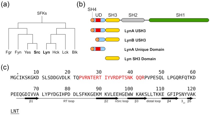
Figure 1.
Schematic representation of Lyn structure. (a) Cladogram representation of the Src Family Kinases (SFKs) according to its evolutionary proximity. (b) Schematic representation of the Lyn protein domains along with the constructs used in the present study. (c) Primary sequence of the LynA USH3 sequence. The natural cysteine in position 3, marked (*), was replaced by serine in the constructs used for chemical shift assignments and chemical shift perturbation. The native cysteine 3 was used to attach a nitroxide probe for paramagnetic relaxation enhancement experiments. Residues absent in Lyn isoform B are shown in red. A schematic representation of the experimentally determined secondary structure elements [13] is presented for the residues in the SH3 domain (E62-T123).

