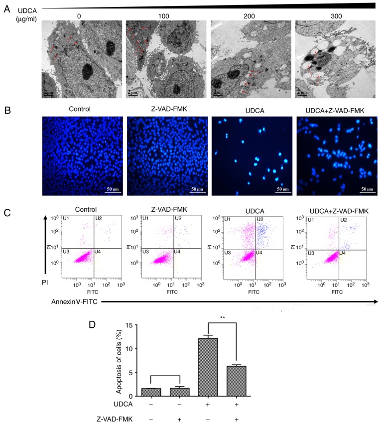
Figure 6.
Transmission electron microscopy to observe morphological changes of mitochondria and the effect of the caspase inhibitor Z-VAD-FMK on apoptotic changes in M14 cells treated with UDCA. Cells were treated with 300 µg/ml UDCA in the presence or absence of 40 µM Z-VAD-FMK. (A) Cells were observed under transmission electron microscopy. Red arrows indicate the location of mitochondria. (B) Morphological apoptotic changes in M14 cells stained with Hoechst 33258. (C) Flow cytometric analysis of apoptosis ratio. (D) The quantification of the apoptosis cell ratio. One representative experiment out of three is presented. **P<0.01. UDCA, ursodeoxycholic acid; PI, propidium iodide; FITC, fluorescein isothiocyanate.

