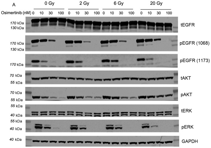
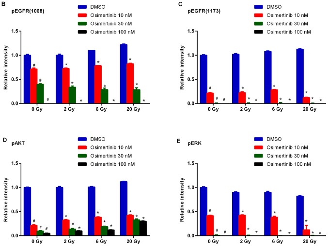
Figure 4.
Western blot analysis of the effect of osimertinib combined with IR on EGFR phosphorylation levels in vitro. Cells were treated with osimertinib 1 h prior to IR, and cell proteins were collected 2 h following IR treatments. GAPDH was included as a loading control. (A) Western blot analysis. (B-E) Quantitative analysis of changes in (B) p-EGFR (1068), (C) p-EGFR (1173), (D) p-AKT and (E) p-ERK, respectively. *P<0.05 vs. IR alone; #P<0.05 vs. DMSO control. IR, irradiation; DMSO, dimethyl sulfoxide; p-, phosphorylated; EGFR, epidermal growth factor receptor; AKT, protein kinase B; ERK, extracellular signal-regulated kinase.


