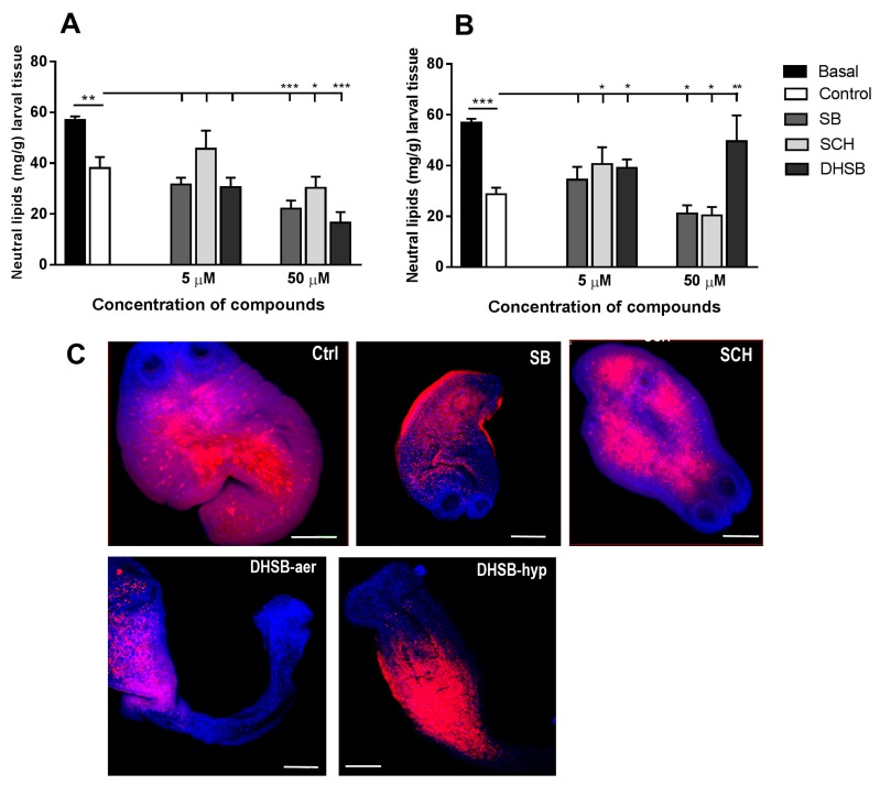
Figure 5.
Neutral lipids concentration in larvae after isolation from mouse (basal), in control group and after in vitro treatment with SB, SCH and DHSB at 5 μM and 50 μM concentrations for 72 h under aerobic (A) and hypoxic (B) conditions. Whole-mount histochemistry and confocal laser scanning microscopy were used to demonstrate the localization of neutral lipids (red) and nuclei (blue) in larvae (C). Images show distribution pattern of lipids in control larvae and after treatment with 50 μM of SB and SCH (upper panel), identical for both aerobic and hypoxic cultivations. Representative images of larvae after DHSB treatment (50 μM, lower panel) showed different staining patterns for individual cultivations. Scale bar = 100 μm, (SB) silybin; (SCH) silychristin; (DHSB) dehydrosilybin. Significantly different values from corresponding controls are shown as * p < 0.05, ** p < 0.01, *** p < 0.001.

