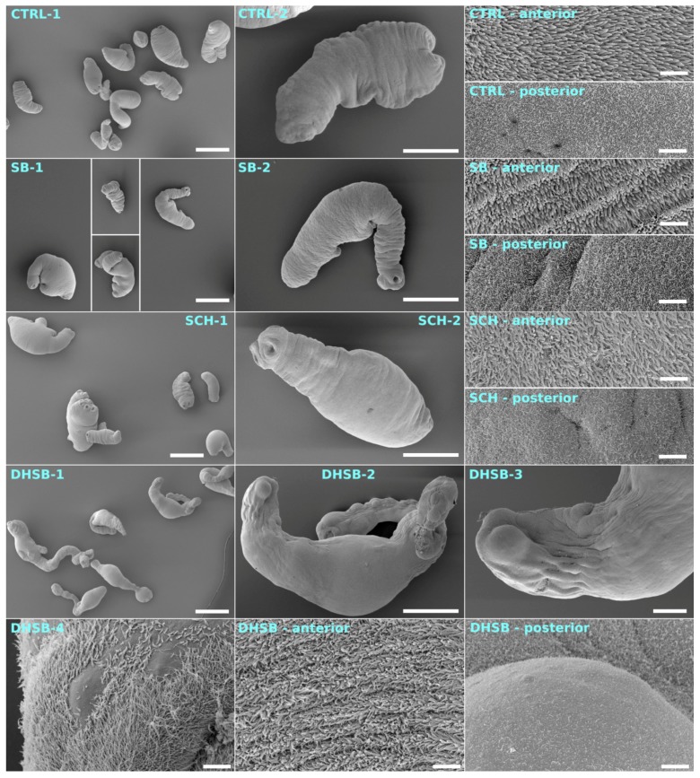
Figure 6.
Effect of 50 μM of SB, SCH and DHSB on whole larval morphology and tegumental alterations after 6 days of incubation were analyzed by scanning electron microscopy. Images placed in left panel (1) and middle panel (2) show shape and morphology of larvae in control (CTRL) and treated groups. Images localized in right and bottom panels represent high resolution microphotographs of tegumental surface on anterior and posterior parts of larvae. Elongation, flattening of larval bodies and narrowing of neck as well as severe damage to tegument seen as aneurysms of tegument and loss of microtriches at posterior part were recorded after DHSB exposure. Scale bars are as follows: CTRL-1, SB-1, SCH-1, DHSB-1—500 μm; CTRL-2, SB-2, SCH-2, DHSB-2—200 μm; DHSB-3—50 μm; DHSB-4—5 μm; all posterior and anterior—5 μm.

