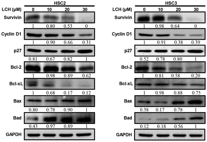
Figure 4.
Effect of LCH on apoptosis regulatory proteins. HSC2 and HSC3 cells were seeded onto a cell culture plate for 24 h, and treated with 10, 20 and 30 µM LCH for 48 h. The proteins were then separated via SDS-PAGE and western blot analysis was performed using survivin, cyclin D1, p27, Bcl-2, Bcl-xL, Bax and Bad antibodies. GAPDH protein was used here as an internal control. LCH, licochalcone H; Bcl-2, B-cell lymphoma 2; Bax, Bcl-2-associated X protein; Bad, Bcl-2-associated death promotor.

