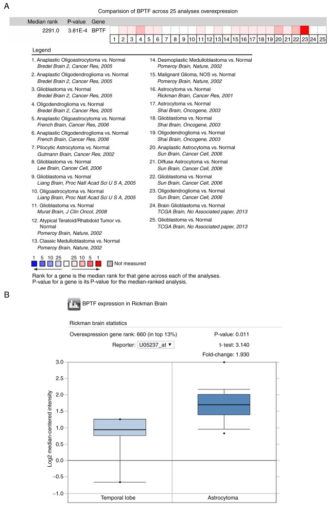
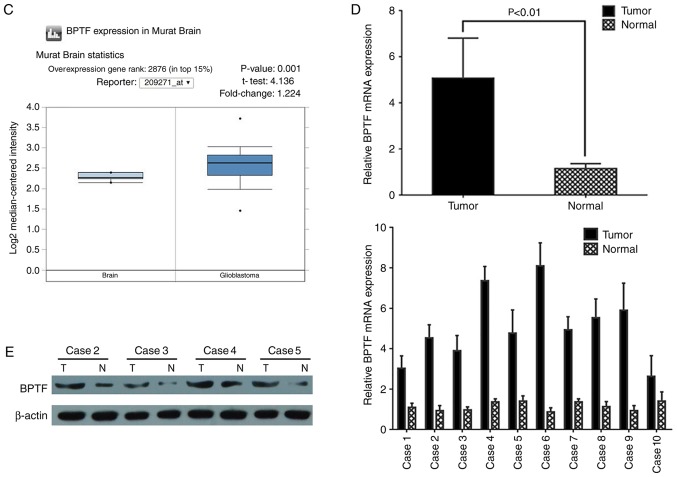
Figure 1.
BPTF expression is significantly elevated in gliomas tissues. (A) The expression profile of BPTF in Oncomine™ database revealed that the expression level of BPTF in brain tumor tissue was significantly higher than that in normal brain tissue in most of the public data. The red color indicated high-rank expression, and the blue color indicated low-rank expression. (B and C) BPTF expression analysis in astrocytoma of Rickman's data (B) (P=0.011) and glioblastoma of Murat's data (C) (P=0.001) were both higher than that in normal brain tissue. (B and C) BPTF expression analysis in astrocytoma of Rickman's data (B) (P=0.011) and glioblastoma of Murat's data (C) (P=0.001) were both higher than that in normal brain tissue. (D) Real-time PCR revealed that BPTF mRNA expression level in glioma tissues was higher than that in corresponding normal brain tissues (P<0.01). (E) The representative western blotting results revealed that BPTF protein expression level in glioma tissues was higher than that in corresponding normal brain tissues.


