Fig. 2.
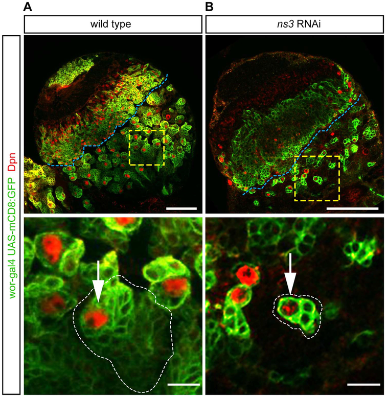
ns3 RNAi mutant NBs in the central brain have reduced lineage sizes. Single slice confocal images of L3 wild type and RNAi brain lobes; NBs are identified by Dpn and NBs and their lineages are marked with GFP (wor-gal4, UAS-mCD8:GFP). (A) Wild type NBs produce large clones of neuronal progeny. Genotype: UAS-Dcr-2; wor-gal4; UAS-mCD8:GFP/UAS-mCherry-RNAi. (B) ns3 RNAi NBs show reduced numbers of neuronal progeny. Genotype: UAS-Dcr-2; wor-gal4; UAS-mCD8:GFP/UAS-ns3-RNAi. The central brain / optic lobe boundary is indicated by a blue line. Representative NBs (white arrows) are boxed in yellow and enlarged in the bottom row. Scale bars: 50 μm top row, 10 μm bottom row.

