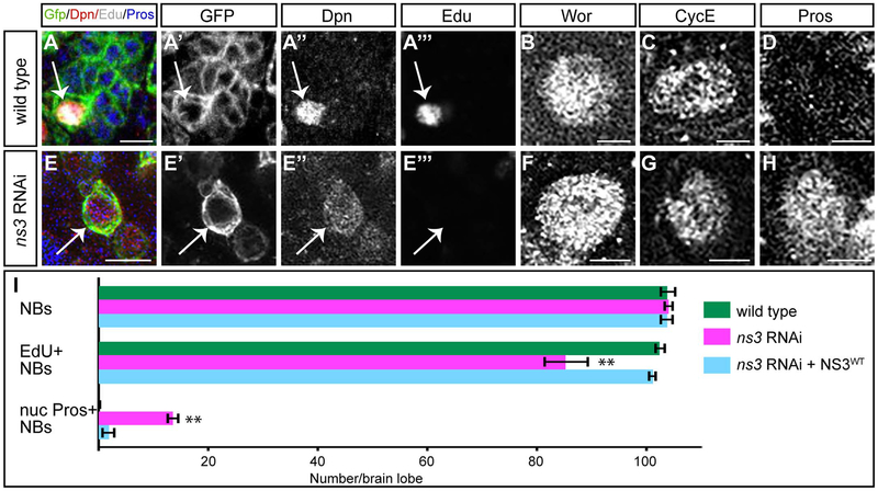
Fig. 3.
Reduced NS3 leads to proliferation arrest in type I NBs. (A-D) Wildtype type I NBs (white arrows) in the L3 brain, identified by wor-gal4 UAS- mCD8:GFP (A′) and Dpn (A”). They can be labeled by a pulse of EdU (A′”), are Wor+ (B) and CycE+ (C), and have no detectable nuclear Pros (D). The Wor, CycE and Pros images were obtained from different confocal stacks. Genotype: UAS-Dcr-2; wor-gal4; UAS-mCD8:GFP/UAS-mCherry-RNAi. (E-H) ns3 RNAi type I NBs (white arrows) in the L3 brain, identified by wor-gal4 UAS- mCD8:GFP (E′) and Dpn (E”). They are not labeled by a pulse of EdU (E′”), but are Wor+ (F) and CycE+(G), and have nuclear Pros (H). The Wor, CycE and Pros images were obtained from different confocal stacks. Genotype: UAS-Dcr-2; wor-gal4; UAS-mCD8:GFP/UAS-ns3-RNAi. (I) Quantifications. n = 5 brain lobes. Error bars indicate s.d. * *: p < 0.001. Scale bars: 10 μm (A, E) and 5 μm (B-D, F-H).

