Fig. 5.
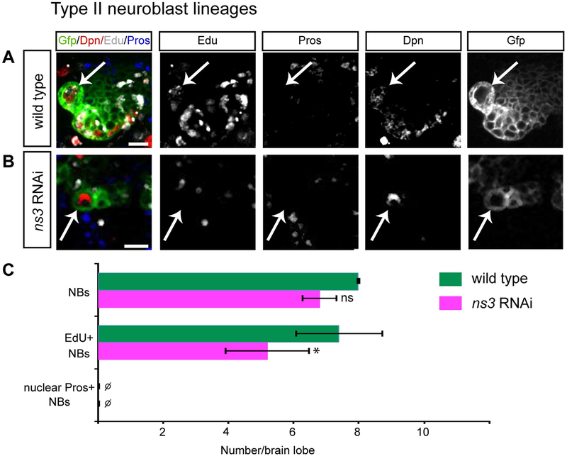
Reduced NS3 leads to proliferation arrest in type II NBs. Type II NBs (white arrows) in the L3 brain, identified by Dpn and wor-gal4 ase-gal80 UAS-GFP. (A) Wild type NBs are labeled by a pulse of EdU. Genotype: UAS-Dcr-2; wor-gal4 ase-gal80; UAS-mCD8:GFP/UAS-mCherry-RNAi. (B) ns3 RNAi NBs are not labeled by a pulse of EdU. Genotype: UAS-Dcr-2; wor-gal4 ase-gal80; UAS-mCD8:GFP/UAS-ns3-RNAi. (C) Quantifications. n = 5 brain lobes. Error bars indicate s.d. *: p < 0.05. Scale bars: 10 μm.

