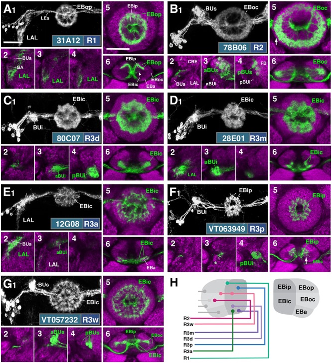
FIGURE 2.

R-neuron subclasses of lineage DALv2/EBa1 with centrifugal arborizations. (A–G) Confocal z-projections of Gal4 drivers that label distinct R-neuron subclasses. Each lettered, six-paneled module corresponds to an individual driver labeled with 10xUAS-mCD8::GFP. Within each module, Top Left (1) is a grayscale z-projection of the specific subclass (cell bodies on left), and its corresponding driver and name in the bottom right corner. Large white annotations designate the primary domains of innervation by the subclass. In remaining module panels, the GFP-labeled neurons are shown in green; neuropil is labeled with anti-DN-cadherin (magenta). Bottom left panels (2–4) are three frontal sections of the bulb at different antero-posterior depths (as described in Figure 1); from left to right: anterior section containing BUa, intermediate section containing aBUs and aBUi, posterior section containing pBUs and pBUi. Some subclasses (R1 and R3a) innervate the LAL rather than the bulb, in which case the same sections are shown at a more ventral position. Top Right (5) is a higher magnification, frontal view of the EB at an antero-posterior level (anterior, intermediate, or posterior) that highlights the circular arbor of a given driver most clearly. Bottom Right (6) is a horizontal section visualizing all five DN-cadherin positive domains. Highlighted in large green text is the domain predominantly innervated by the R-neuron subclass; smaller green text signifies additional regions of innervation. Small white text in all panels denotes relevant spatial landmarks. (A1–6) R31A12-Gal4 (R1). (A1–5) Refer to Supplementary Movie 1. (A1) R1 projects from the LAL to EBop. For all DALv2 R-neurons, the anterior component of the lateral ellipsoid fascicle (LEa) comprises the bridge between proximal and distal, annular neurites. (A2–4) Ventral neurites of R1 extend in the lateral LAL, medially adjacent of the gall (GA). (A5) Posterior EB section. (A6; refer to Supplementary Movie 2) R1 neurites in the EB line the anterior-most border of EBop, along the EBip–EBop interface. Additional, very small protrusions emanate from canal projections, along the EBip-EBic interface (arrowhead; A6). (B1–6) R78B06-Gal4 (R2). (B1–5) Refer to Supplementary Movie 3. (B1) R2 projects from BUs to EBoc. (B2) No significant innervation in BUa; GFP signal corresponds to bypassing neurites. (B3) R2 neurons exhibit most of their microglomeruli in aBUs, but (B4) also some in pBUs. (B5) Intermediate EB section; arrow indicates peripheral fringe of EBoc which is not innervated. (B6; refer to Supplementary Movie 4) R2 exhibits restricted innervation of EBoc; GFP signal in EBic is passing neurites. (C1–6) R80C07-Gal4 (R3d – distal). (C1–5) Refer to Supplementary Movie 5. (C1) R3d projects from BUi to EBic. (C2) No significant innervation in BUa; GFP signal corresponds to bypassing neurites. (C4) R3d neurons exhibit most of their microglomeruli in pBUi, but (C3) also some in aBUi. (C5) Intermediate EB section. (C6; refer to Supplementary Movie 6) R3d fills most of EBic. (D1–6) R28E01-Gal4 (R3m – medial). (D1–5) Refer to Supplementary Movie 7. (D1) R3m projects from BUi to EBic. (D3) R3m neurons exhibit most of their microglomeruli in aBUi, but (D2) possibly also extend fibrous projections in the LAL, adjacent to BUa. (D4) No significant innervation in pBUs/i. (D5) Anterior EB section. (D6; refer to Supplementary Movie 8) R3m fills complementary region of EBic relative to R3d. (E1–6) R12G08-Gal4 (R3a – anterior). (E1–5) Refer to Supplementary Movie 9. (E1) R3a projects from the LAL to EBic. (E2) The LAL projections of R3a neurons are more closely adjacent to BUa, than those of R1 neurons (A2), and (E3) may also exhibit very sparse projections in aBUi. (E4) No significant innervation in pBUs/i. (E5) Anterior EB section. (E6; refer to Supplementary Movie 10) The EB neurites of R3a surround EBa. (F1–6) VT063949-Gal4 (R3p – posterior). (F1–5) Refer to Supplementary Movie 11. (F1) R3p projects from BUi to EBip. (F2,3) No significant innervation in BUa and aBUs/i; GFP signal corresponds to bypassing neurites. (F4) R3p neurons exhibit their microglomeruli in pBUi. (F5) Posterior EB section. R3p neurites in the EB densely fill EBip, but (F6; refer to Supplementary Movie 12) also appear to project anteriorly, penetrating EBic (arrow). (G1–6) VT057232-Gal4 (R3w – wide). (G1–5) Refer to Supplementary Movie 13. (G1) R3w projects from BUs to EBic. (G2) No significant innervation in BUa; GFP signal corresponds to bypassing neurites. (G3,4) R3w neurons exhibit their microglomeruli in aBUs and pBUs. (G5) Posterior EB section. (G6; refer to Supplementary Movie 14) R3w neurites line the posterior border of EBic, and extend into EBip. Neurites also extend distally toward EBoc, and may encroach on it. (H) Schematized overview of EB domain innervation patterns of centrifugally projecting R-neurons. CRE, crepine. Scale bars represent 25 μm (A–G).
