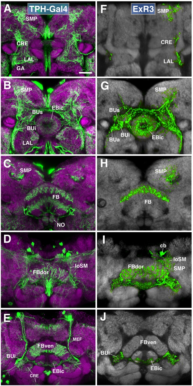
FIGURE 7.

ExR3-neurons: posterior extrinsic ring neurons of lineages DPMpm2/DM3. (A–C,E) Panel format similar to Figures 5B–E. Confocal z-projections of the TPH-Gal4 driver that labels all serotonergic neurons, including ExR3-neurons. (D) Additional row depicting dorsal section of the FB (FBdor). (F–J) Confocal z-projections of individually labeled cells generated by MCFO using TPH-Gal4. Five panels depict the same sections and are organized in the same fashion as in panels A–E. Neuropil labeled by anti-DN-cadherin (gray). An individual ExR3 neuron innervates (F–H) the LAL, the crepine (CRE) and SMP in a predominantly ipsilateral fashion, but (F,G) also sparsely innervates the same compartments contralaterally. (G) Additional innervation includes and all BU partitions on both sides, along with EBic. (H,I) ExR3 exclusively innervates the dorsal strata of the FB. (I) Dorsal view illustrates that the cell body (cb) of an individual cell, located in the dorso-posterior cortex, sends a projection along the longitudinal superior medial fascicle (loSM) and arborizes in panel (J) EBic. CRE, crepine; FB, fan-shaped body; FBven, ventral part of the fan-shaped body; MEF, medial equatorial fascicle; ML, medial lobe of the mushroom body; NO, noduli; SMP, superior medial protocerebrum. Scale bars represent 25 μm (A–J).
