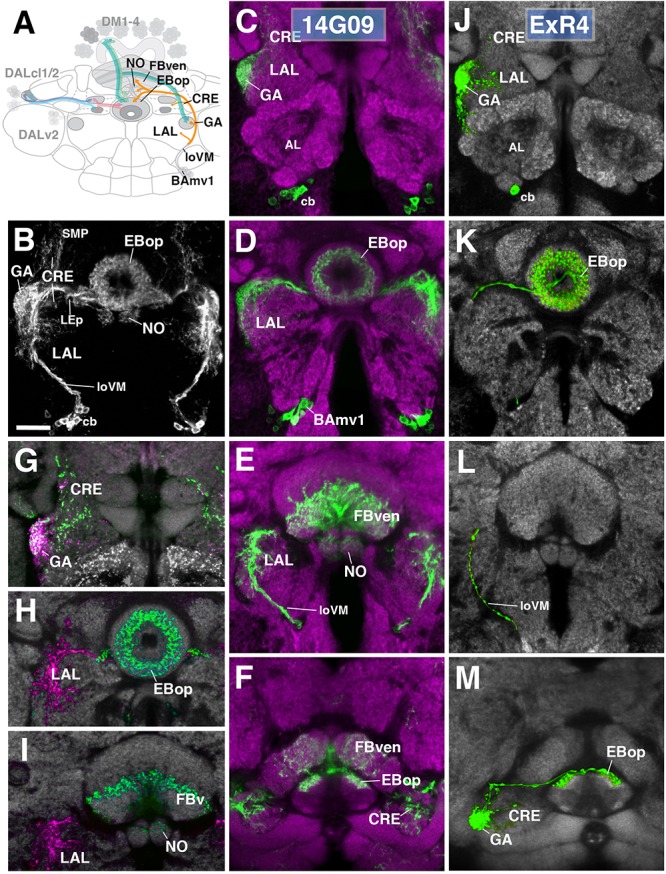
FIGURE 8.

Anterior ExR-neuron subclass of lineage BAmv1/LALv1. (A) Schematized overview of interacting neuronal populations of the EB from Figure 1, now including anterior ExR-neurons of lineage BAmv1 (orange). (B) Grayscale z-projection of ExR4, labeled by R14G09 > 10xUAS-mCD8::GFP. Z-projection spans from the gall (GA)/lateral accessory lobe (LAL) to the EB, and does not include the FB component of this driver (see below). Cell bodies (cb) of BAmv1 neurons send projections dorsally along the medial component of the ventral longitudinal fascicle (loVM), which then extend from the GA/LAL to EBop. (C–F) Top three rows corresponds to frontal sections of R14G09 > 10xUAS-mCD8::GFP at three different antero-posterior depths (refer to Supplementary Movie 23). From top to bottom: (C) anterior section containing the GA/LAL, (D) intermediate section containing the EB and bulbs, (E) posterior section containing the FB and noduli (NO). Bottom row (F; refer to Supplementary Movie 24) is a horizontal section visualizing the length of the EB canal. R14G09-positive neurons are shown in shown in green; neuropil is labeled with anti-DN-cadherin (magenta). (G–I) Corresponding sections in panels C,D using R14G09 labeled with the presynaptic marker syt.EGFP (green) and dendritic marker DenMark (red). Neuropil is labeled with anti-DN-cadherin (gray). (J–M) Confocal z-projections of individually labeled cells generated by MCFO using R14G09-Gal4. Four panels depict the same sections and are organized in the same fashion as in panels C–F. Neuropil labeled by anti-DN-cadherin (gray). An individual ExR4 neuron, with (J) its cell body (cb) located ventrally of the antennal lobe (AL), predominantly innervates the GA, with fine processes into the LAL, and (K) EBop. (L) ExR4 projects along the loVM and does not innervate the FB. (M) Dorsal view illustrates the innervation in the GA and arborization in EBop. CRE, crepine; FBven, ventral region of the FB; LEp, posterior component of the lateral ellipsoid fascicle; SMP, superior medial protocerebrum. Scale bars represent 25 μm (B–M).
