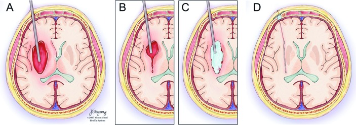
Figure 2.
(A) The endoscope sheath is inserted 1.5 cm from the distal wall of the hematoma under stereotactic guidance. (B) Blood is aspirated until the brain closes in around the end of the sheath at which point it is retracted 1–2 cm. (C) When the sheath retracts to the proximal wall of the hematoma cavity, saline is infused through the sheath, inflating the hematoma cavity. (D) After evacuation, the sheath is removed, and burr hole ultrasound followed by DYNA CT are performed to confirm adequate removal.

