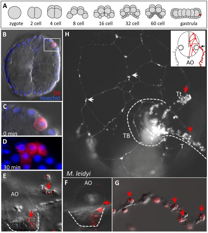
Fig. 7.
Neurons and colloblasts share a common progenitor. (A) Embryonic cleavage stages in Mnemiopsis leidyi. Random micromeres (red) were injected at the late gastrula stage. (B) Optical section of a live gastrula immediately following injection of DiI (red); nuclei are labeled with Hoechst. (C) High magnification image of the boxed area in B showing a single injected micromere. (D) High magnification image of a clone of labeled cells 30 min after DiI injection. (E–G) A live cydippid larva with two labeled populations of cells: in the floor plate of the apical organ (AO) and the colloblasts of the tentacle (T). (E) Both cell populations are depicted in a single focal plane. (F) A different focal plane showing labeling of cells and dome cilia (arrow) on one side of the apical organ. (G) High magnification image of the extended tentacle; colloblasts (arrows) are the only labeled cells in the tentacle. (H) Cydippid larva with DiI-labeled neurons of the subepidermal nerve net in the body wall (white arrows) and colloblasts (red arrows); the animal is viewed from the tentacular plane (black arrow) as summarized in the inset. TB—tentacle bulb, T—tentacle, Tt—tentilla.

