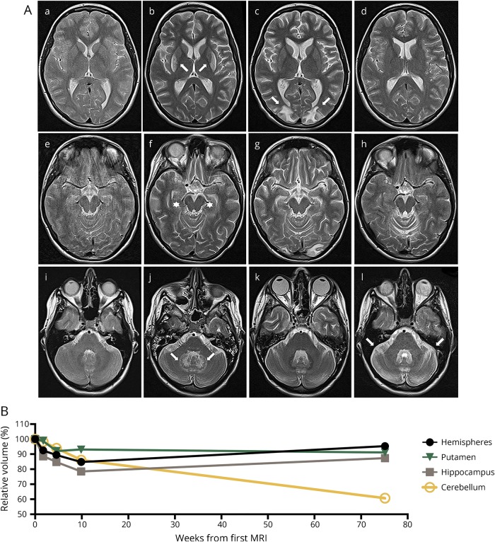
Figure. Multiple MRI abnormalities as correlates of diverse adverse events and neurologic impairment during the course of severe NMDAR-E.
(A) Transaxial T2w MRIs at levels depicting the basal ganglia (A.a–A.d), the mesiotemporal structures (A.e–A.h), and the cerebellum (A.i–A.l) are shown. MRI at initial presentation showed no abnormalities (A.a, A.e, and A.i). After 72 days of isoflurane, T2w signal hyperintensities in the lateral striatum and the deep cerebellar nuclei developed (A.b and A.j, arrows). Temporomesial atrophy became evident (A.f, stars). Remission of the T2w signal hyperintensities after discontinuation of isoflurane while after therapy-refractory arterial hypertension, PRES with typical vasogenic edema developed in bilateral occipital regions areas (A.c, arrows). Mesiotemporal atrophy proceeded. Nine months after disease onset, signal abnormalities associated with PRES were in full remission while severe cerebellar atrophy had occurred (A.l, arrows). (B) MRI volumetry performed using the FMRIB Software Library (FSL, Version 5.0.9, fmrib.ox.ac.uk/fsl, for details see ref. 13) demonstrates a mesiotemporally pronounced reversible atrophy hardly affecting the putamen and progressive cerebellar atrophy. Volumes of the different structures were normalized to the volume of the initial MRI. NMDAR-E = NMDA receptor encephalitis; PRES = posterior reversible encephalopathy syndrome; T2w = T2-weighted.

