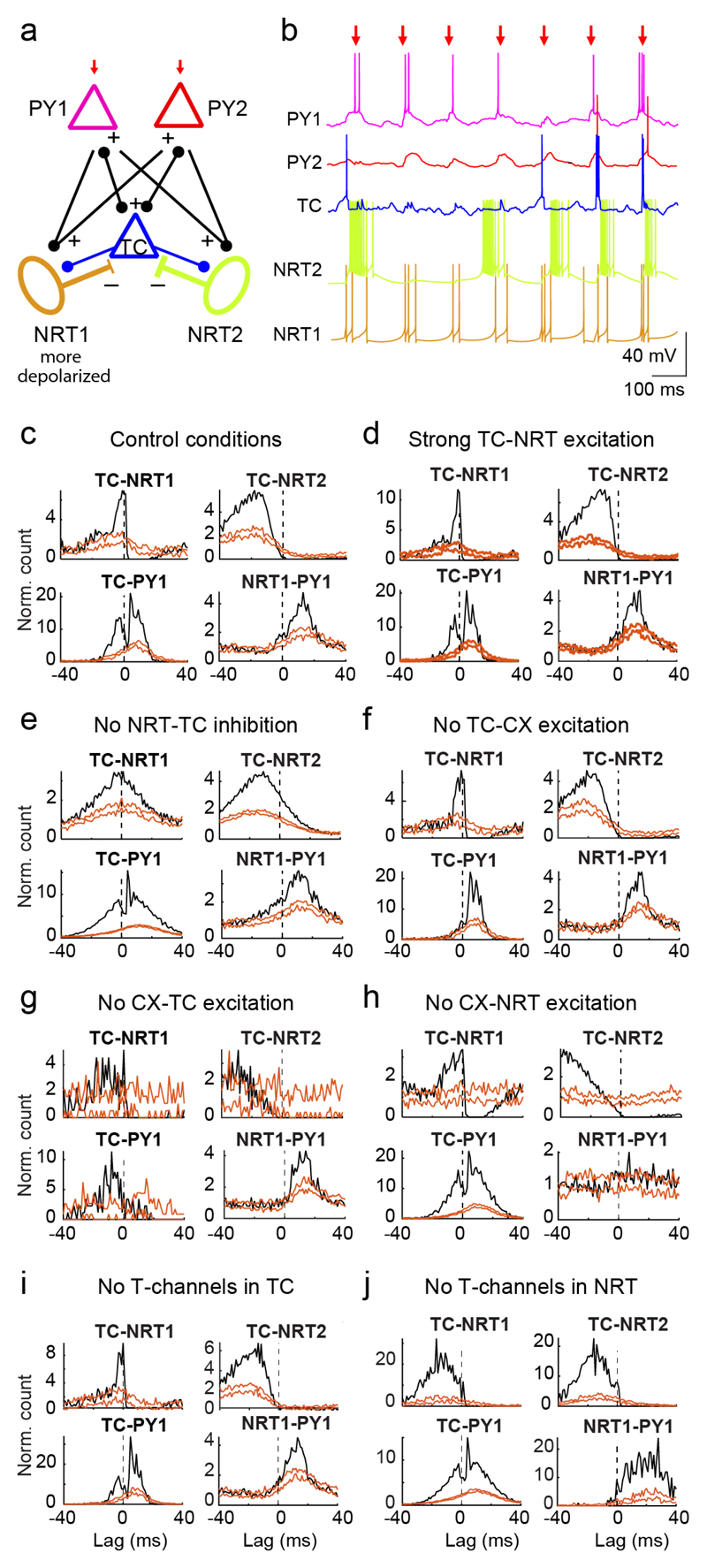
Figure 8. Firing interactions in a cortico-thalamic network model.
(a) Schematic diagram of the model network, consisting of 1 TC, 2 NRT and 2 excitatory cortical pyramidal neurons (PY). NRT1 is more depolarized than NRT2 via steady injected current. Excitatory (AMPA) synapses connect TC to NRT and PY neurons, and PY neurons to NRT and TC neurons. Inhibitory GABAA synapses connect NRT to TC neurons. Only PY neurons receive a train of 5 AMPA EPSPs that is repeated at a frequency of 7 Hz (red arrows indicate the 3rd EPSP in the train which was used as time-reference for the analysis). (b) Example membrane potential traces illustrate the firing dynamics during 7 Hz stimulation protocol. Note the presence of high frequency bursts in NRT2 and their paucity in TC and NRT1 neuron. (c-j) XCors (black) of simulated firing of different neuron pairs under various conditions (orange lines indicate expected confidence intervals (5-95%) estimated as in Fig. 5 using the 3rd EPSPs as time-reference). The simulation under “control” conditions well reproduced the experimental XCors (c), and no major change was observed in simulations with an increase in the strength of the TC-to-NRT neuron synapse up to 6-fold that of the CX-NRT neuron synapses (d), whereas blocking the NRT-to-TC neuron GABAA synapses abolished the trough after time zero in TC-NRT neuron XCors (e). Block of cortical to TC neuron synapses or vice versa led to TC-CX XCors that were different from those observed experimentally (f, g), and while a trough was still present in TC-NRT XCors when the cortical input to NRT neurons was absent, there was no peak in NRT-CX XCors (h). Removal of T-channels in TC neurons had no major effect on the XCors (i), whereas in NRT neurons led to broader XCors peaks (j) compared to the control conditions (a).

