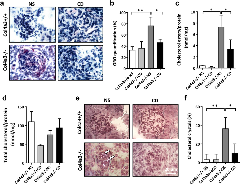
Figure 3. Effect of HPβCD on cholesterol homeostasis in Col4a3 knockout mice.
(A, B) Representative oil red O (ORO) staining of kidney sections (4 μm) (A) and bar graph analysis (B) from wildtype mice treated with normal saline (Col4a3+/+ NS) or cyclodextrin (Col4a3+/+ CD) and Col4a3 knockout mice treated with normal saline (Col4a3−/− NS) or HPβCD (Col4a3−/− CD) (40x). Bars are 20μm. *p<0.05, **p<0.01, One-way ANOVA. (C) Bar graph analysis showing significant reduction of cholesterol esters accumulation in Col4a3 knockout mice after 4 weeks of HPβCD treatment. *p<0.05, One-way ANOVA. (D) Bar graph analysis showing that HPβCD treatment does not affect total cholesterol levels in kidney cortexes of Col4a3 knockout mice when compared to wild type littermates. (E, F) Cholesterol crystal staining (E) and bar graph analysis (F) indicating significant cholesterol crystal accumulation in Col4a3 knockout mice compared to wild type littermates. HPβCD treatment leads to significant reduction in cholesterol crystals in Col4a3 knockout mice. Bars are 20μm. *p<0.05, **p<0.01, One-Way ANOVA. All data are presented as mean ± SD.

