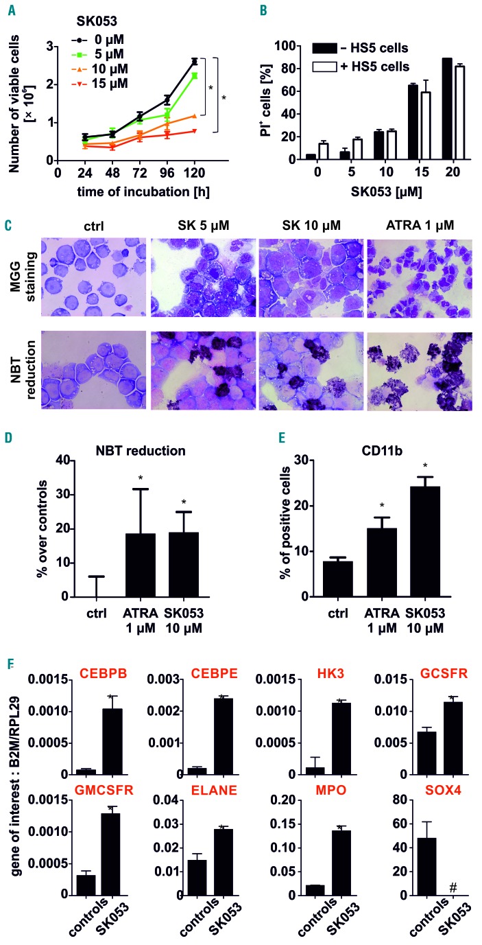
Figure 1.
SK053 induces differentiation of AML cells. (A) HL-60 cells were incubated with various concentrations of SK053 for up to 5 days. Each day the number of dead cells was evaluated with trypan blue exclusion (mean ± SD from 6 experiments). (B) Co-culture of HL-60 cells with universal bone marrow stromal cells (HS5) did not impair SK053 cytotoxicity. HL-60 cells were seeded in a transwell system on top of HS5 cells and incubated with SK053 for 72 h. Next, HL-60 cells were analyzed for viability using propidium iodide (PI) staining in flow cytometry (data are shown as mean percentages of PI-positive cells ± SD from 3 experiments); *P<0.05 in one-way ANOVA with the Dunnett post-hoc test. (C) May-Grünwald-Giemsa (MGG) staining (upper panel) and nitroblue tetrazolium (NBT) (lower panel) in HL-60 cells incubated with SK053 for 120 h. All-trans retinoic acid (ATRA) served as a positive control. (D) Semi-quantitative colorimetric NBT reduction assay in HL-60 cells incubated for 5 days with 10 mM SK053 (data are shown as mean increases, in %, over controls ± SD for 3 experiments); *P<0.05 in one-way ANOVA with the Dunnett post-hoc test. (E) Mean percentage ± SD of HL-60 cells expressing CD11b myeloid marker was determined in flow cytometry in 3 experiments; *P<0.05 in one-way ANOVA with the Dunnett post hoc test. (F) Real-time quantitative polymerase chain reaction analysis of HL-60 cells incubated for 5 days with 10 μM SK053; results are presented as mean target-to-reference ratio ± SD of three experiments; *P<0.05 vs. controls, two-tailed Student t test, #P<0.05 vs. controls, one-tailed Student t test.

