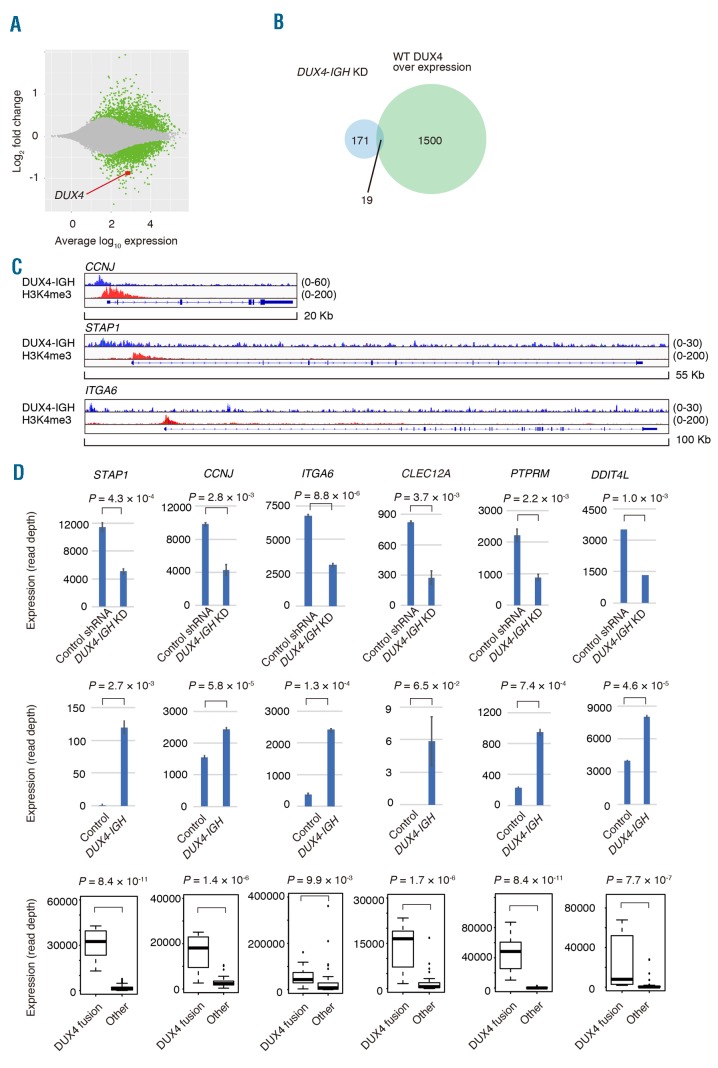
Figure 2.
Knockdown of DUX4-IGH with short hairpin RNA. (A) MA plot showing the change in the transcripts with DUX4-IGH knockdown. Genes with a significant change (P<0.05) in expression are plotted in green, and DUX4 is plotted in red. (B) Venn diagram showing the number of genes underrepresented in DUX4-IGH knockdown in NALM6 cells (light blue) or genes whose expression was upregulated by overexpression of wild-type (WT) DUX4 in NALM6 cells (light green). (C) Mapped reads at the CCNJ, STAP1, or ITGA6 locus obtained with chromatin immunoprecipitation coupled with sequencing (ChIP-seq) against DUX4-IGH (blue) or trimethylated lysine residue at position four of histone H3 (H3K4me3, red) in NALM6 cells. (D) Expression of representative genes in NALM6 infected with control shRNA or shRNA against DUX4-IGH (upper panel). Expression of the genes in Reh infected with control vector or DUX4-IGH expression vector (middle panel). Expression of the genes in clinical samples of B-ALL with or without DUX4 fusion (lower panel).

